Mesoscopic Light Transport Properties of a Single
Biological Cell and Ultra-Early Cancer Detection
Quantification of Nanoscale Density Fluctuations in Biological Cells/Tissues by Electron Microscopy Imaging During Early Carcinogenesis
Inverse Participation Ratio (IPR) study of TEM iamages of cells and tissues
(Optical IPR Imaging of Nano-Architecture of a Single
Biological Cells/Tissues with length scale variations )
Most cancers are curable if they are diagnosed and treated at an early stage. Recent studies suggest that nanoarchitectural changes occur within cells during early
carcinogenesis which precede microscopically evident tissue changes. It follows that the ability to comprehensively interrogate cell nanoarchitecture (e.g., macromolecular
complexes, DNA, RNA, proteins, and lipid membranes) could be critical to the study of early carcinogenesis. We have developed a technique to study the nanoscale
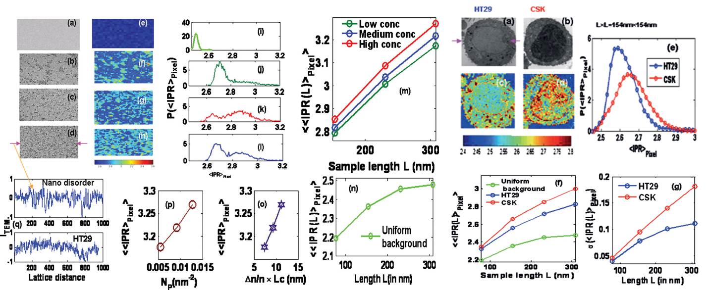
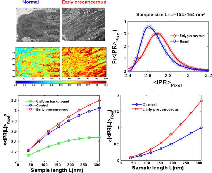
mass-density fluctuations of biological cells and tissues by quantifying their nanoscale light-localization properties. Transmission electron microscope (TEM) images of
human colon cancer cell line (HT29) variants and tissues are used to construct corresponding effective disordered optical lattices. Light-localization properties are
studied by statistical analysis of the inverse participation ratio (IPR) of the eigenfunctions of these optical lattices at the nanoscale. The IPR analysis is validated in
experiments with models of disordered systems fabricated from dielectric nanoparticles. Experimental results from genetically modified HT29 cell line variants show a
statistically significant increase of the average IPR value, which parallels the genetic events leading to an increase in the aggressive growth of these cell lines. A
similar trend of IPR values is shown in rectal tissues obtained from human subjects diagnosed with an early stage colon neoplasia. These results indicate an elevation of
the nanoscale disorder strength (e.g., refractive index fluctuations) of cells in the field of early carcinogenesis. The IPR technique provides the only early-stage
detection technique of nanoarchitectural alterations in carcinogenesis known to date and could be a potential means of characterizing cells and tissues.
Publications
-
"Quantification of Nanoscale Density Fluctuations using Electron Microscopy:
Light Localization Properties of Biological Cells"
Prabhakar Pradhan, D. Damania, H. Joshi, V. Turzhitsky, H. Subramanian,
H. K. Roy, A. Taflove, V. P. Dravid, V. Backman
Applied Physics Letters 97 , 243704 (2010)
Link to the journal /
PDF
-
Quantification of nanoscale density fluctuations by electron microscopy: probing cellular
alterations in early carcinogenesis
Prabhakar Pradhan, D. Damania, H. Joshi, V. Turzhitsky, H. Subramanian,
H. K. Roy, A. Taflove, V. P. Dravid, V. Backman
Physical Biology 8, 026012 (2011)
Link to the journal /
PDF
You are visitor #
 since April 1, 2005,
Return
since April 1, 2005,
Return